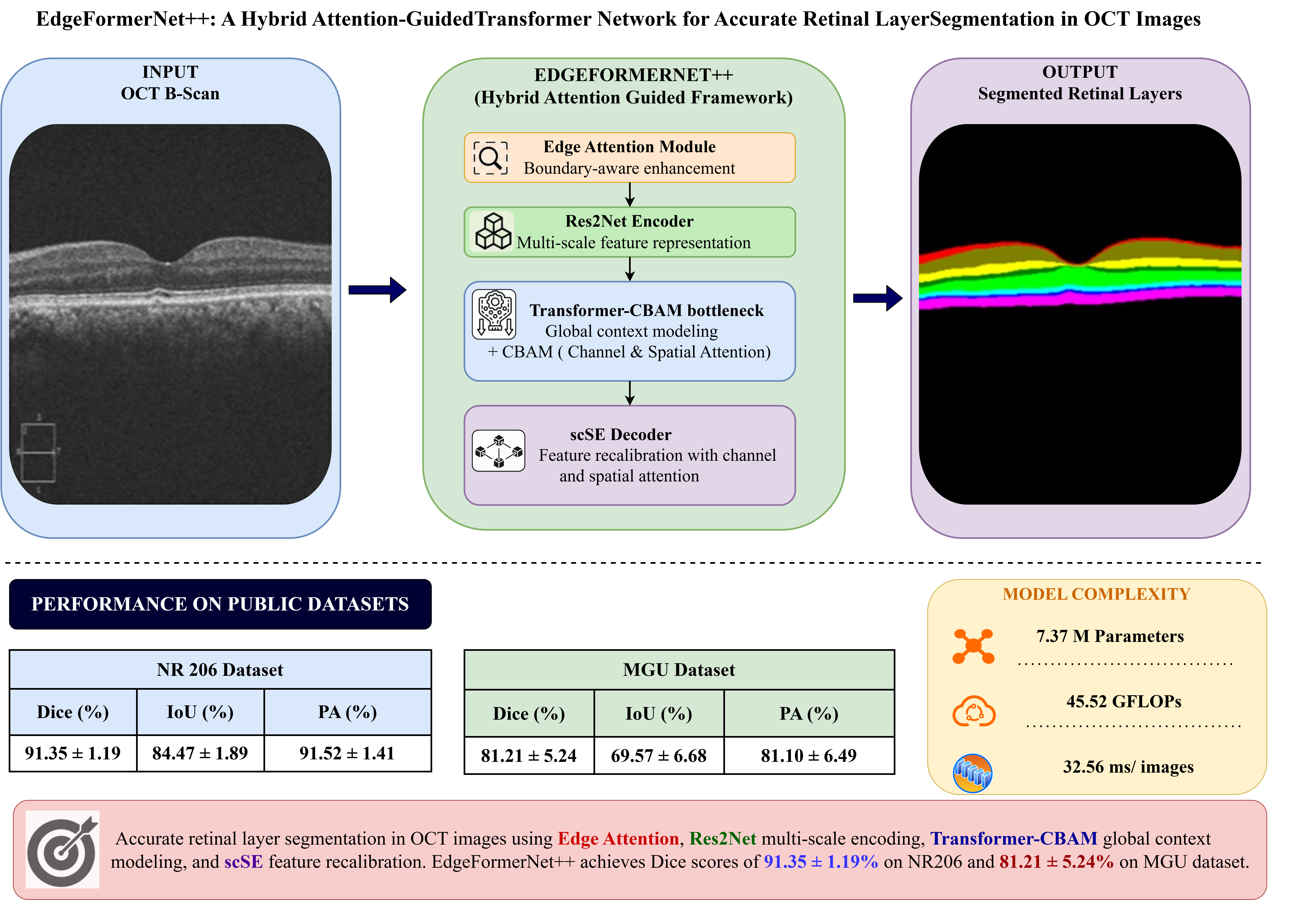
EdgeFormerNet++: A Hybrid Attention-Guided Transformer Network for Accurate Retinal Layer Segmentation in OCT Images
Keywords:
Deep Learning, Edge Attention, Hybrid CNN-Transformer Architecture, Multi-class Semantic Segmentation, Optical Coherence Tomography (OCT), Res2Net, Retinal Layer Segmentation.Abstract
Accurate retinal layer segmentation in OCT is crucial for the early detection and monitoring of retinal and neurodegenerative diseases, yet it remains challenging due to the thin, low-contrast structures and closely packed layers in multi-class settings. We propose EdgeFormerNet++, a hybrid framework that integrates Res2Net-based multi-scale encoding, lightweight Transformer bottlenecks for global context, and dual attention (CBAM and scSE) for spatial-channel recalibration, complemented by an edge attention module to refine layer boundaries. The model is evaluated on two public datasets (NR206 and MGU) covering macular and peripapillary regions, achieving state-of-the-art Dice scores of 92.34% and 83.28%, respectively. Qualitative and quantitative results demonstrate accurate delineation of complex retinal structures, supporting the use of EdgeFormerNet++ for automated OCT analysis and computer-assisted diagnosis.
Downloads
References
I. La´ıns, J. C. Wang, et al., “Retinal applications of swept source optical coherence tomography (oct) and optical coherence tomography angiography (octa),” Progress in Retinal and Eye Research, vol. 84, p. 100 951, 2021. DOI: 10 . 1016 / j . preteyeres . 2021 .100951.
J. Nathans, “Seeing is believing: The development of optical coherence tomography,” Proceedings of the National Academy of Sciences, vol. 120, no. 39, e2311129120, 2023. DOI:10.1073/pnas.2311129120.
A. London, I. Benhar, and M. Schwartz, “The retina as a window to the brain—from eye research to cns disorders,” Nature Reviews Neurology, vol. 9, no. 1, pp. 44–53, 2013. DOI: 10.1038/nrneurol.2012.227.
C. Rascun`a, A. Russo, et al., “Retinal thickness and microvascular pattern in early parkinson’s disease,” Frontiers in Neurology, vol. 11, p. 533 375, 2020. DOI: 10.3389/fneur.2020.533375.
H. Xie et al., “Arterial hypertension and retinal layer thickness: The beijing eye study,” British Journal of Ophthalmology, vol. 108, no. 1, pp. 105–111, 2024. DOI: 10.1136/bjo-2022-322229.
O. Ronneberger, P. Fischer, and T. Brox, “U-net: Convolutional networks for biomedical image segmentation,” in International Conference on Medical Image Computing and Computer-Assisted Intervention, Springer, 2015, pp. 234–241. DOI: 10.1007/978-3-319-24574-4_28.
Z. Zhang, Q. Liu, and Y. Wang, “Road extraction by deep residual u-net,” IEEE Geoscience and Remote Sensing Letters, vol. 15, no. 5, pp. 749–753, 2018. DOI: 10.1109/LGRS.2018.2802944.
O. Oktay et al., “Attention u-net: Learning where to look for the pancreas,” in Medical Image Computing and Computer-Assisted Intervention – MICCAI 2018, vol. 11070, Springer, 2018, pp. 121–130. DOI: 10 .48550/arXiv.1804.03999.
H. Cao et al., “Swin-unet: Unet-like pure transformer for medical image segmentation,” in Computer Vision – ECCV 2022 Workshops, Springer, 2022, pp. 205–218.
DOI: 10.1007/978-3-03125066-8 9.
S. Jardim, J. Ant´onio, and C. Mora, “Image thresholding approaches for medical image segmentation — short literature review,” Procedia Computer Science, vol. 219, pp. 1485–1492, 2023. DOI: 10.1016/j.procs.2023.01.439.
S. Pare, A. Kumar, G. K. Singh, and V. Bajaj, “Image segmentation using multilevel thresholding: A research review,” Iranian Journal of Science and Technology, Transactions of Electrical Engineering, vol. 44, no. 1, pp. 1–29, 2020. DOI: 10.1007/s40998-019-00251-1.
J. Liu et al., “Automated retinal boundary segmentation of optical coherence tomography images using an improved canny operator,” Scientific Reports, vol. 12, no. 1, p. 1412, 2022. DOI: 10.1038/s41598-022-05550-y.
Y. Chen, P. Ge, G. Wang, G. Weng, and H. Chen, “An overview of intelligent image segmentation using active contour models,” Intelligent Robotics, vol. 3, no. 1, pp. 23–55, 2023. DOI: 10.20517/ir.2023.02.
K. Ramesh, G. K. Kumar, K. Swapna, D. Datta, and S. S. Rajest, “A review of medical image segmentation algorithms,” EAI Endorsed Transactions on Pervasive Health & Technology, vol. 7, no. 27, 2021. DOI: 10.4108/eai.12-4-2021.169184.
M. A. Chandra and S. S. Bedi, “Survey on svm and their application in image classification,” International Journal of Information Technology, vol. 13, no. 5, pp. 1– 11, 2021. DOI: 10.1007/s41870-017-0080-1.
T. Zhu, “Analysis on the applicability of the random forest,” in Journal of Physics: Conference Series, vol. 1607, IOP Publishing, 2020, p. 012 123. DOI: 10.1088/1742-6596/1607/1/012123.
R. Azad et al., “Medical image segmentation review: The success of u-net,” IEEE Transactions on Pattern Analysis and Machine Intelligence, vol. 46, no. 12, pp. 10 076–10 095, 2024. DOI:10.1109/TPAMI.2024.3435571.
A. G. Roy et al., “Relaynet: Retinal layer and fluid segmentation of macular optical coherence tomography using fully convolutional networks,” Biomedical Optics Express, vol. 8, no. 8, pp. 3620–3637, 2017. DOI: 10.1364/BOE.8.003627.
B. M. Zipporah and D. F. X. Christopher, “Residual u-net architecture for retinal layers in oct images with choroidal neovascularization,” International Journal of Electrical and Electronics Engineering, vol. 10, no. 5, pp. 205–212, 2023. DOI: 10.14445/23488379/IJEEEV10I5P119
B. Fazekas et al., “Sd-layernet: Semi-supervised retinal layer segmentation in oct using disentangled representation with anatomical priors,” in Medical Image Computing and Computer-Assisted Intervention – MICCAI 2022 Springer, 2022, pp. 320–329. DOI: 10.1007/978-3-031-16452-1_31.
J. Chen et al., “Transunet: Rethinking the u-net architecture design for medical image segmentation through the lens of transformers,” Medical Image Analysis, vol. 97, p. 103 280, 2024. DOI: 10.1016/j.media.2024.103280.
Y. Gao, M. Zhou, and D. N. Metaxas, “Utnet: A hybrid transformer architecture for medical image segmentation,” in Medical Image Computing and Computer-Assisted Intervention – MICCAI 2021,
Springer, 2021, pp. 61–71. DOI: 10.1007/978-3-030-87199-4 6.
A. Hatamizadeh et al., “Unetr: Transformers for 3d medical image segmentation,” in Proceedings of the IEEE/CVF winter conference on applications of computer vision, 2022, pp. 574–584. DOI: https://doi.org/10.48550/arXiv.2103.10504.
J. Li et al., “Multi-scale gcn-assisted two-stage network for joint segmentation of retinal layers and discs in peripapillary oct images,” Biomedical Optics Express, vol. 12, no. 4, pp. 2204–2220, 2021. DOI: 10 . 1364 /BOE.417212.
E. Liu et al., “Mt net: A multi-scale framework using the transformer block for retina layer segmentation,”Photonics, vol. 11, p. 607, 2024. DOI: 10 . 3390 /photonics11070607.
X. He et al., “Lightweight retinal layer segmentation with global reasoning,” IEEE transactions on instrumentation and measurement, vol. 73, pp. 1–14, 2024. DOI: 10.1109/TIM.2024.3400305.
J. Hao, H. Li, S. Lu, Z. Li, and W. Zhang, “General retinal layer segmentation in oct images via reinforcement constraint,” Computerized Medical Imaging and Graphics, vol. 120, p. 102 480, 2025. DOI: 10.1016/j.compmedimag.2024.102480.
H. Peng, Y. Yu, and S. Yu, “Re-thinking the effectiveness of batch normalization and beyond,” IEEE transactions on pattern analysis and machine intelligence, vol. 46, no. 1, pp. 465–478, 2023. DOI: 10.1109/TPAMI.2023.3319005.
K. Hara, D. Saito, and H. Shouno, “Analysis of function of rectified linear unit used in deep learning,” in 2015 international joint conference on neural networks (IJCNN), IEEE, 2015, pp. 1–8. DOI: 10.1109/IJCNN.2015.7280578.
S.-H. Gao, M.-M. Cheng, K. Zhao, X.-Y. Zhang, M.-H. Yang, and P. Torr, “Res2net: A new multi-scale backbone architecture,” IEEE transactions on pattern analysis and machine intelligence, vol. 43, pp. 652–662, 2019. DOI: 10.1109/TPAMI.2019.2938758.
S. Woo, J. Park, J.-Y. Lee, and I. S. Kweon, “Cbam: Convolutional block attention module,” in Proceedings of the European Conference on Computer Vision (ECCV), 2018, pp. 3–19. DOI: 10.1007/978- 3- 030-01234-2_1.
A. G. Roy, N. Navab, and C. Wachinger, “Recalibrating fully convolutional networks with spatial and channel ‘squeeze & excitation’ blocks,” in Medical Image Computing and Computer-Assisted Intervention – MICCAI 2018, vol. 11071, Springer, 2018, pp. 733–741. DOI:10.1109/TMI.2018.2867261.
P. Gholami, P. Roy, and V. Lakshminarayanan, “Octid: Optical coherence tomography image database,” Data in Brief, vol. 28, p. 104 882, 2020. DOI: 10 . 1016 / j .compeleceng.2019.106532.
A. Buslaev, V. I. Iglovikov, E. Khvedchenya, A. Parinov, A. Druzhinin, and A. A. Kalinin, “Albumentations: Fast and flexible image augmentations,” Information, vol. 11, no. 2, p. 125, 2020. DOI: 10 . 3390 /info11020125.
F. Milletari, N. Navab, and S.-A. Ahmadi, “V-net: Fully convolutional neural networks for volumetric medical image segmentation,” in Proceedings of the Fourth International Conference on 3D Vision (3DV), IEEE, 2016, pp. 565–571. DOI: 10.1109/3DV.2016.79.
O. Ronneberger, P. Fischer, and T. Brox, “U-net: Convolutional networks for biomedical image segmentation,” in Medical Image Computing and Computer- Assisted Intervention (MICCAI), vol. 9351, Springer, 2015, pp. 234–241. DOI: 10.1007/978-3-319-24574-4_28.
F. Isensee, P. F. Jaeger, S. A. A. Kohl, J. Petersen, and K. H. Maier-Hein, “Nnu-net: A self-configuring method for deep learning-based biomedical image segmentation,” Nature Methods, vol. 18, no. 2, pp. 203–211, 2021. DOI: 10.1038/s41592-020-01008-z.
X. He et al., “Exploiting multi-granularity visual features for retinal layer segmentation in human eyes,” Frontiers in Bioengineering and Biotechnology, vol. 11, p. 1 191 803, 2023. DOI: 10.3389/fbioe.2023.1191803.